Research Assistant Boston Children's Hospital Boston, Massachusetts
Abstract:
Purpose: Supplemental bone grafting is often required prior to dental implant placement in patients with cleft lip and palate (CLP) even after successful alveolar bone grafting (ABG).1 Many surgical techniques have been described to augment the alveolar ridge and prepare it for implant placement but there is little consensus on which approach yields the most predictable results.2 The purpose of this study was to evaluate whether onlay cortical block bone grafting at lateral incisor sites achieves sufficient bone for implant placement in patients with CLP.
Patients and
Methods: Retrospective case series of patients with CLP who had cortical block bone grafting for placement of a dental implant to replace a missing lateral incisor(s) at Boston Children’s Hospital from 2015 through 2020. Patients were excluded if follow up details regarding implant placement were absent, CBCT scans were unavailable or quality of scans insufficient for performing our measurements. The primary predictor was onlay cortical block grafting to lateral incisor site either harvested from the mandibular ramus or symphysis. Primary outcome was change in alveolar bone width at edentulous lateral incisor site 1.) 3 mm apical to the adjacent central incisor cementoenamel junction (CEJ) and 2.) 1 mm above the apex of adjacent central incisor as measured on pre- and post-operative cone beam computed tomography (CBCT) scans. Covariates included age, gender, cleft type (unilateral vs. bilateral), and time between grafting and post-operative CBCT. Secondary outcomes included need for further bone augmentation and implant placement. Data analyses was performed using Pearson X2 and Student’s t-test. Intrarater reliability was measured by intra-class correlation coefficients (ICC) using a two-way mixed model calculated for absolute agreement. A P-value of < 0.05 was considered statistically significant
Results: A total of 11 lateral incisor sites were evaluated (mean age 18.9 (range 2.2) years, 44% male). Six patients had unilateral CLP and 3 patients had a bilateral CLP. One site was grafted with a symphysis block graft and the remaining 10 sites were treated with ramus grafting. The mean width of the alveolar ridge on post-operative CBCT after grafting was 6.19 mm 3 mm apical to adjacent central incisor CEJ (point A) and 9.38 mm 1 mm above adjacent central incisor root apex (point B). At point A, patients experienced a mean (standard deviation) increase in bone width of 3.03mm (1.80). At point B, there was a mean (standard deviation) change in bone width of 3.39mm (1.12). The change in bone width was statistically significant at both levels (p < 0.001, p< 0.001). Age, gender, cleft type (unilateral vs. bilateral), and time between graft and post-operative CBCT were not associated with differences in the amount of bone augmentation achieved. All lateral incisor sites had implants placed with successful osseointegration and only one site required additional grafting at the time of implant placement. The ICCs for all measurements were greater than 0.9, indicating excellent intrarater agreement.
Conclusions: Cortical block onlay bone grafting is an acceptable technique to augment lateral incisor sites in patients with cleft lip and palate for placement of a dental implant.
References: 1. Green MA, Padwa BL. Does Timing of Secondary Alveolar Bone Grafting Affect the Need for Additional Bone Augmentation Prior to Implant Placement at Cleft Sites? J Oral Maxillofac Surg. 2021 Sep;79(9):1927-1931. 2. Jensen SS, Terheyden H. Bone augmentation procedures in localized defects in the alveolar ridge: clinical results with different bone grafts and bone-substitute materials. Int J Oral Maxillofac Implants. 2009;24 Suppl:218-36.
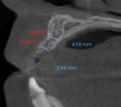
Pre operative measurements taken prior to onlay cortical block bone grafting at edentulous alveolar cleft lateral incisor site.
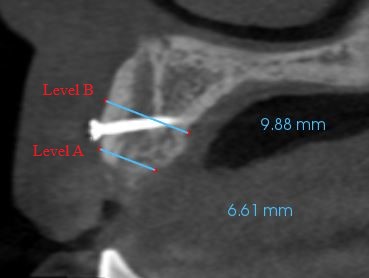
Post operative measurements taken after onlay cortical block bone grafting at edentulous alveolar cleft lateral incisor site